
Cell Organelles and Their Functions
Cellular life hinges on intricate organelle interactions, potentially unlocking disease understanding like cancer and neurodegeneration. Advanced imaging reveals organelle movement and novel discoveries.
Cell organelles are specialized subunits within a cell, each performing a distinct function crucial for the cell’s survival and overall operation. These microscopic structures, found within eukaryotic cells, are analogous to organs in a complex organism, working in harmony to maintain life. Understanding these components is fundamental to comprehending biological processes.
Recent research highlights the importance of studying interactions within and between organelles, suggesting these connections hold keys to understanding complex diseases. The ability to directly visualize how newly formed organelles transition within living cells, thanks to advances in imaging, is revolutionizing our understanding. The discovery of previously unknown organelles further emphasizes the dynamic and complex nature of the cellular world.
Essentially, these membrane-bound structures carry out various functions, and their coordinated activity is essential for life. Investigating these organelles opens doors to potential treatments for inherited diseases and a deeper understanding of cellular mechanisms.
The Cell: A Basic Unit of Life
The cell stands as the fundamental building block of all known living organisms, representing the smallest unit capable of performing life functions. Trillions of these microscopic units compose the human body, each a bustling hub of activity orchestrated by specialized structures – organelles. A cell’s core components include the membrane, nucleus, and cytoplasm, each playing a vital role in maintaining cellular integrity and function.

Understanding the cell’s intricate organization is paramount, as it provides insight into how organisms develop, function, and respond to their environment. Recent discoveries emphasize that the interactions between organelles are critical, potentially unlocking solutions to complex diseases. The ability to visualize organelle movement and identify novel structures underscores the cell’s dynamic nature.
Ultimately, the cell’s functionality relies on the coordinated efforts of its organelles, making their study essential for advancing biological knowledge and medical treatments.
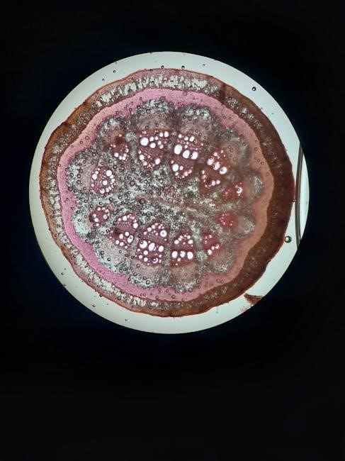
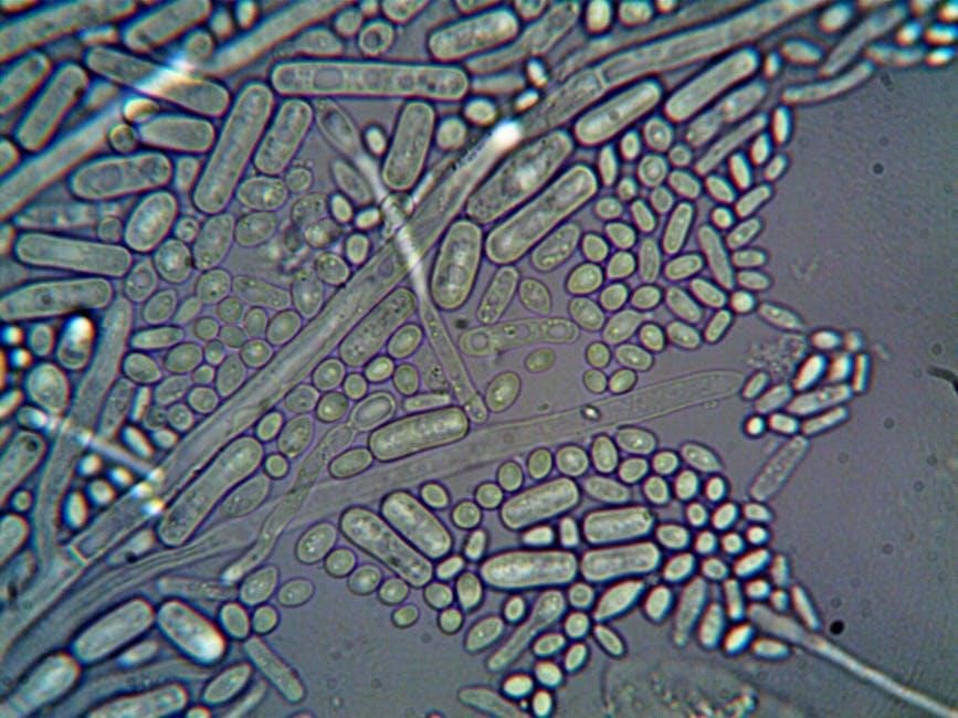
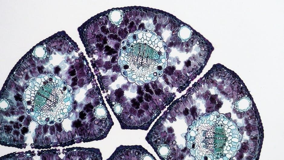
Prokaryotic vs. Eukaryotic Cells
Cells broadly fall into two categories: prokaryotic and eukaryotic, distinguished by their structural complexity. Prokaryotic cells, found in bacteria and archaea, lack a nucleus and other membrane-bound organelles. Their genetic material resides in the cytoplasm. Conversely, eukaryotic cells, comprising plants, animals, fungi, and protists, possess a defined nucleus housing their DNA and a diverse array of internal organelles.
This distinction is crucial; eukaryotic cells exhibit a higher degree of organization, with organelles performing specialized functions. The presence of these organelles allows for compartmentalization, enhancing efficiency and complexity. Understanding these differences is vital, as it impacts how cells function and interact with their environment.
Recent research highlights the importance of organelle interactions within eukaryotic cells, potentially revealing insights into disease mechanisms. The study of these cellular structures is fundamental to biological understanding.

Major Cell Organelles and Their Roles
Eukaryotic cells contain little, membrane-bound organelles that carry out diverse functions, essential for life’s processes and cellular health within complex organisms.
Nucleus: The Control Center
The nucleus reigns supreme as the cell’s command center, safeguarding and meticulously regulating genetic material – DNA. This vital organelle dictates cellular activities, growth, and reproduction. Encased by a double membrane known as the nuclear envelope, it provides a secure environment for the genome.
Within the nucleus, DNA isn’t simply a tangled mess; it’s organized into chromosomes. These structures become visible during cell division. The nucleus also houses the nucleolus, a region dedicated to ribosome biogenesis – the creation of essential protein-building machinery.
Essentially, the nucleus controls protein synthesis by transcribing DNA into RNA, which then exits the nucleus to guide protein production. Its integrity is paramount; damage to the nucleus can lead to cellular dysfunction and potentially, diseases like cancer. Understanding the nucleus is fundamental to comprehending life itself.
Endoplasmic Reticulum (ER): Synthesis and Transport
The Endoplasmic Reticulum (ER) forms an extensive network of membranes throughout the cytoplasm, serving as the cell’s manufacturing and transport hub. This dynamic organelle plays a crucial role in synthesizing lipids, proteins, and steroids, alongside the crucial task of calcium storage. Recent imaging reveals how newly formed organelles transition from the ER onto microtubule tracks.
The ER exists in two primary forms: rough and smooth, each with specialized functions. Its interconnectedness allows for efficient movement of molecules throughout the cell. The ER’s ability to modify and fold proteins ensures their proper function, while its transport capabilities deliver these proteins to their designated locations.
Essentially, the ER is a cellular highway and factory combined, vital for maintaining cellular homeostasis and responding to changing conditions. Its proper function is essential for overall cell health and survival.
Rough Endoplasmic Reticulum (RER)
The Rough Endoplasmic Reticulum (RER) is characterized by ribosomes attached to its outer surface, giving it a “rough” appearance. These ribosomes are the sites of protein synthesis, making the RER central to the production of proteins destined for secretion, insertion into membranes, or localization within other organelles.
As proteins are synthesized, they enter the RER lumen where they undergo folding and modification. This ensures correct protein structure and function. The RER also plays a role in quality control, identifying and degrading misfolded proteins to prevent their accumulation.
The RER’s extensive network facilitates efficient protein transport throughout the cell, delivering proteins to the Golgi apparatus for further processing and packaging. It’s a critical component in the cellular protein production and quality control pathway.
Smooth Endoplasmic Reticulum (SER)
The Smooth Endoplasmic Reticulum (SER) lacks ribosomes, giving it a smooth appearance, and performs diverse metabolic functions. Unlike the RER, its primary role isn’t protein synthesis, but rather lipid and steroid hormone synthesis. It’s heavily involved in creating and storing these vital molecules.
The SER also participates in carbohydrate metabolism, specifically glycogen breakdown in liver cells, and detoxification of drugs and poisons. This detoxification process often involves modifying harmful substances to make them more water-soluble for excretion.
Furthermore, the SER stores calcium ions, crucial for muscle contraction and signaling in nerve cells. Its tubular network extends throughout the cell, contributing to efficient transport and distribution of these essential compounds, showcasing its versatility.
Golgi Apparatus: Processing and Packaging
The Golgi apparatus, a central component of the endomembrane system, functions as a molecular warehouse and finishing factory for the cell. It receives proteins and lipids from the endoplasmic reticulum, further processing and modifying them for specific destinations.
This organelle consists of flattened, membrane-bound sacs called cisternae, arranged in stacks. As molecules move through these stacks, they undergo glycosylation – the addition of carbohydrate chains – which is vital for protein folding and function.
The Golgi then sorts and packages these modified molecules into vesicles, small membrane-bound sacs, for transport to other organelles or secretion outside the cell. This precise packaging ensures proteins reach their correct locations, highlighting its crucial role in cellular logistics.
Mitochondria: Powerhouse of the Cell
Mitochondria are often hailed as the “powerhouses” of the cell, and rightfully so. These double-membrane-bound organelles are responsible for generating most of the cell’s supply of adenosine triphosphate (ATP), the primary energy currency.
Through a process called cellular respiration, mitochondria break down glucose and other fuel molecules, releasing energy that is then captured in ATP. The inner mitochondrial membrane is highly folded into cristae, increasing the surface area for ATP production.
Interestingly, mitochondria possess their own DNA, separate from the cell’s nuclear DNA, suggesting an ancient symbiotic origin. Their function extends beyond energy production, playing roles in signaling, cellular differentiation, and even programmed cell death, making them vital for overall cellular health.
Lysosomes: Waste Management and Digestion
Lysosomes function as the cell’s primary waste disposal and digestive system. These membrane-bound organelles contain a potent cocktail of enzymes capable of breaking down a wide variety of cellular debris, including worn-out organelles, macromolecules, and even invading pathogens.
Through a process called autophagy, lysosomes engulf and digest damaged or unnecessary cellular components, recycling their building blocks. They also participate in phagocytosis, where they engulf external materials like bacteria.
Maintaining a proper pH within the lysosome is crucial for optimal enzyme activity. Dysfunction of lysosomes can lead to the accumulation of undigested materials, contributing to various inherited diseases. Their role in cellular cleanup is essential for maintaining cellular homeostasis and preventing harmful build-ups.
Organelles Involved in Synthesis and Transport
Ribosomes and vacuoles are key players, facilitating protein creation and storage, alongside crucial transport mechanisms within the cellular environment for optimal function.
Ribosomes: Protein Synthesis
Ribosomes are fundamental cellular structures responsible for protein synthesis, a process vital for all life forms. These aren’t membrane-bound organelles, existing freely in the cytoplasm or attached to the endoplasmic reticulum. They translate genetic code from messenger RNA (mRNA) into amino acid sequences, building proteins essential for cellular functions.
The process begins when mRNA binds to a ribosome. Transfer RNA (tRNA) molecules, each carrying a specific amino acid, recognize corresponding codons on the mRNA. As the ribosome moves along the mRNA, tRNA delivers amino acids, which are linked together to form a polypeptide chain. This chain then folds into a functional protein.
Ribosomes come in two subunits – large and small – which combine during translation. Their efficiency and accuracy are crucial for maintaining cellular health. Errors in protein synthesis can lead to dysfunctional proteins and contribute to various diseases. Understanding ribosome function is therefore paramount in biological research.
Vacuoles: Storage and Support
Vacuoles are membrane-bound cell organelles with diverse functions, primarily involved in storage and maintaining cell turgor pressure. Found in both plant and animal cells, their size and content vary significantly depending on the cell type and its needs. In plant cells, a large central vacuole occupies a substantial volume, storing water, nutrients, and waste products.
This central vacuole also plays a crucial role in maintaining cell rigidity, providing support against the cell wall. In animal cells, vacuoles are typically smaller and involved in storing and transporting materials, as well as waste removal. They can also participate in processes like autophagy, breaking down and recycling cellular components.
Essentially, vacuoles act as dynamic storage compartments, adapting to the cell’s changing requirements. Their ability to regulate cell volume and internal pressure is vital for overall cell health and function.
Emerging Research and Novel Organelles
Recent discoveries reveal previously unknown organelles, potentially revolutionizing treatments for inherited diseases. Imaging techniques are crucial for understanding complex organelle interactions.
Peroxisomes: Detoxification
Peroxisomes are vital microbodies found in nearly all eukaryotic cells, playing a crucial role in metabolic processes, particularly detoxification. These single-membrane bound organelles are characterized by their ability to break down various molecules, including fatty acids, through oxidation reactions.
A key enzyme within peroxisomes is catalase, which decomposes hydrogen peroxide – a harmful byproduct of metabolic activities – into water and oxygen. This detoxification function is essential for protecting the cell from oxidative damage. Beyond detoxification, peroxisomes also participate in the synthesis of certain lipids, like plasmalogens, important components of cell membranes.

Furthermore, they contribute to the breakdown of excess purines and the metabolism of specific amino acids. Dysfunction of peroxisomes can lead to severe genetic disorders, highlighting their indispensable role in cellular health and overall organismal well-being. Ongoing research continues to unveil the complexities of peroxisomal function and their involvement in diverse cellular pathways.
Cytoskeleton: Structural Support and Movement
The cytoskeleton is a dynamic network of protein filaments extending throughout the cytoplasm of eukaryotic cells, providing essential structural support and facilitating cellular movement. Composed of three primary types – microtubules, actin filaments, and intermediate filaments – it’s far from a static scaffold.
Microtubules, crucial for intracellular transport and cell division, act as tracks for motor proteins. Actin filaments are vital for cell shape, motility, and muscle contraction. Intermediate filaments offer tensile strength and anchor organelles. This intricate network isn’t just about structure; it’s actively remodeled to respond to cellular signals.
Recent imaging reveals how newly formed organelles utilize microtubule tracks for transport, demonstrating the cytoskeleton’s dynamic role. Disruptions in the cytoskeleton are linked to various diseases, emphasizing its importance. Understanding its function is key to comprehending cellular processes and disease mechanisms.
Cell Membrane: Boundary and Communication
The cell membrane, a vital boundary, isn’t merely a passive barrier; it’s a dynamic interface controlling what enters and exits the cell. Composed primarily of a phospholipid bilayer with embedded proteins, it maintains cellular integrity while enabling crucial communication with the external environment.

These embedded proteins function as channels, transporters, receptors, and markers, facilitating selective permeability and signal transduction. The membrane’s fluidity allows for flexibility and adaptation, essential for processes like endocytosis and exocytosis. It’s a constantly shifting mosaic, responding to cellular needs.
Effective communication relies on the membrane’s ability to receive and transmit signals. Disruptions in membrane function can lead to cellular dysfunction and disease. Understanding its structure and function is fundamental to comprehending cellular life and its interactions with the world.
Recent Discoveries of Unknown Organelles
The cellular landscape is continually evolving, with recent breakthroughs revealing previously unknown organelles within our cells. These specialized structures, often membrane-bound, challenge existing understandings of cellular organization and function. One such discovery, dubbed a novel organelle, holds potential for new treatments targeting devastating inherited diseases.
Researchers are actively investigating the precise roles of these newly identified compartments, exploring their involvement in various cellular processes. These discoveries highlight the limitations of current models and emphasize the need for continued exploration.
The identification of these organelles underscores the complexity of the cell and opens exciting avenues for research. Further investigation promises to unveil their mechanisms and potential therapeutic applications, revolutionizing our approach to disease treatment.
Imaging Techniques and Organelle Interactions
Advancements in imaging technologies are revolutionizing our understanding of how organelles interact within the dynamic cellular environment. For the first time, scientists can directly visualize the departure of newly formed organelles from the endoplasmic reticulum (ER) and their subsequent travel along microtubule tracks inside living cells.
These real-time observations provide crucial insights into the mechanisms governing organelle trafficking and communication. Sophisticated microscopy techniques allow researchers to track organelle movements, assess their proximity, and identify key protein interactions.
Such detailed visualizations are essential for deciphering the complex interplay between organelles and understanding how disruptions in these interactions contribute to disease development; This is crucial for understanding complex diseases.
Organelle Dysfunction and Disease
The intricate functions of cell organelles are fundamental to health, and their dysfunction is increasingly linked to a wide range of diseases. Disruptions in organelle interactions, as revealed by advanced imaging, can contribute to the development of complex conditions like cancer and neurodegenerative disorders.
The discovery of previously unknown organelles suggests that our understanding of cellular processes is incomplete, and defects in these structures may underlie devastating inherited diseases. Investigating these novel organelles could unlock new therapeutic targets.
Understanding how organelle malfunction contributes to disease pathogenesis is crucial for developing effective treatments. Targeting specific organelle pathways may offer a promising avenue for intervention and disease management, improving patient outcomes.